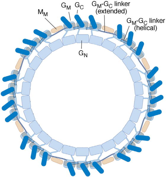
Fig. 8.

Model for the organization of FliG subunits in the flagellar switch complex. The view is from the top (the membrane-proximal side). The three domains of FliG, and the middle domain of FliM, are shown. Copy numbers of FliG and FliM vary somewhat from specimen to specimen [46]; the complex pictured has 26 FliG and 34 FliM subunits. Each FliGM domain (present in 26 copies) rests on a FliMM domain (present in a 34-fold array). To accommodate the copy-number mismatch while allowing every FliGC to stack onto the FliGM domain of an adjacent subunit, the segment linking FliGM to FliGC takes on an extended rather than helical conformation at the gap positions.
