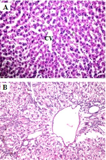
Figure 3.

Histopathological Examination of Liver. (A) Normal liver histology of a rat fed the standard diet. (B) Rats administrated DEN display focal area of anaplastic hepatocytes with other cells forming acini were observed associated with fibroblastic cells proliferation dividing the degenerated and necrosed hepatic parenchyma into nodules (H and E X400).
