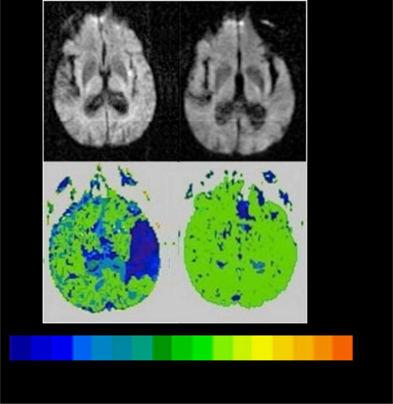
Figure 4.

Scans showing reperfusion of left pSTG associated with auditory word comprehension,
Top Panel. DWI on Day 1(left) and Day 3 (right) showing tiny area of infarct in left insular cortex.
Lower Panel. PWI on Day 1 (left) and Day 3 (right) showing severe hypoperfusion of left superior pSTG on Day 1 and reperfusion of left superior pSTG on Day 3, when auditory word comprehension had recovered. The color bar shows the number of seconds of delay in time to peak arrival of contrast, compared to normally perfused tissue (0 seconds).
