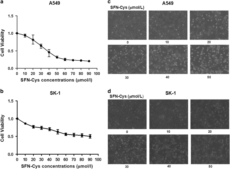
Figure 1.
Cell viability analysis and morphological changes after treated with SFN-Cys. (a) A549 and (b) SK-1 cells were treated with various concentrations of SFN-Cys (0, 10, 20, 30, 40, 50, 60, 70 and 80 μM) for 24 h. SFN-Cys significantly inhibited the proliferation of A549 and SK-1 cells in a dose-dependent manner. These results are presented as mean±S.D. from three independent experiments. Error bars indicate uncertainty errors in graphs to relay 95% of confidence in the interpreted data. The (c) A549 and (d) SK-1 cells were examined with Leica DMIRB Microscope at ×100 magnification. Cells treated with various concentrations of SFN-Cys (0, 10, 20, 30, 40 and 50 μM) generated morphological changes.

