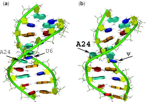
Figure 2.
A schematic view of uBP (a) and ψBP (b) from structures solved by Newby and Greenbaum (8; uBP PDB:1LMV; ψBP PDB: 1LPW. Model #1 from both duplexes was used). RNA helices were rendered using the DINO (http://www.dino3d.org) visualization program. The color schemes are as follows: the backbone is green; the sugar is yellow; base A is cyan; base C is red; base G is orange and base U and ψ are blue. Note that uBP adopts a typical A-helical pattern, whereas ψBP is characterized by an extrahelical branch site adenosine.

