Fig. 2.
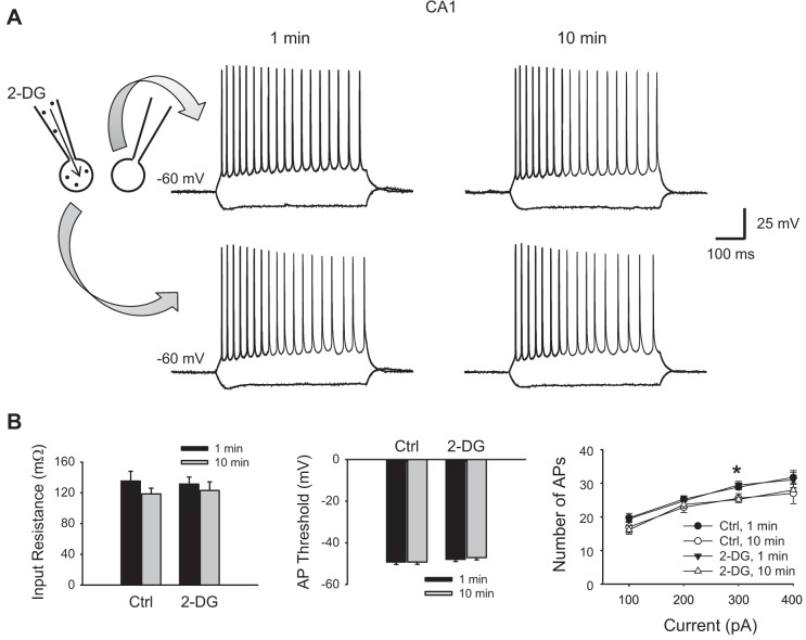
Glycolytic inhibition with 2-DG does not alter basal membrane excitability of hippocampal CA1 neurons. A: two neighboring CA1 pyramidal cells were recorded concurrently, one with 10 mM intracellular 2-DG and one as a control without 2-DG. Their membrane responses to current injection pulses (−200 to +500 pA, 100-pA step, 500 ms) right after rupture of the cell (~1 min) and 10 min after rupture are shown at left and right, respectively. Note both neurons displayed a regular firing pattern. B: summary data showing that the membrane input resistance (left) and action potential (AP) threshold (middle) were not different between the 2-DG-loaded neurons and controls and did not change over time in the presence of intracellular 2-DG (P > 0.05, ANOVA, n = 7 pairs). Right, input/output (I/O) relationship (i.e., number of APs as a function of current injection) was also similar between the 2-DG-loaded and control neurons but was slightly reduced after 10 min, particularly at one point in the larger current range (300 pA, *P < 0.05). However, both 2-DG-loaded and nonloaded neurons changed in the same direction, and there was no significant difference between them (P > 0.05, ANOVA, followed by Holm-Sidak test).

