Figure 3.

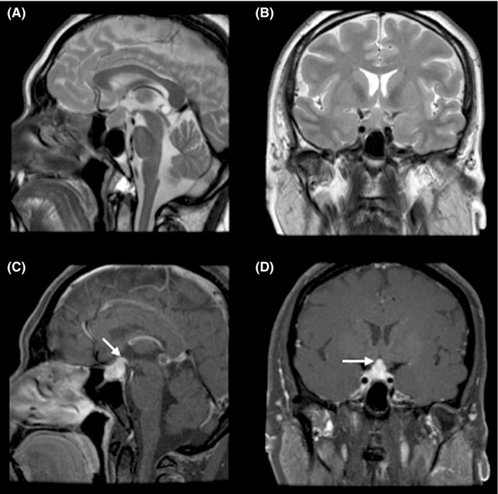
MRI scan of the brain, axial view (A) and coronal view T2WI (B) showing isointense sellar, suprasellar lesion. (C&D) after intravenous injection of gadolinium, showing enhancement of the mass lesion.

MRI scan of the brain, axial view (A) and coronal view T2WI (B) showing isointense sellar, suprasellar lesion. (C&D) after intravenous injection of gadolinium, showing enhancement of the mass lesion.