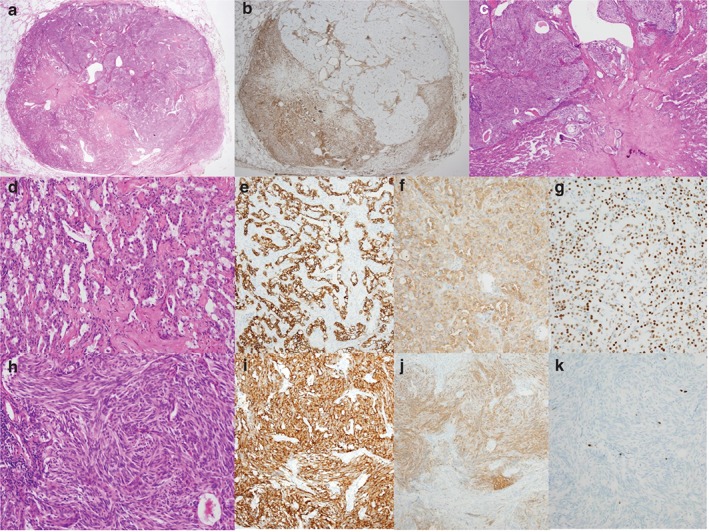
Figure 3.

Microscopic findings of the postoperative specimen. (a) Hematoxylin and eosin (H&E) stain shows a well‐circumscribed non‐encapsulated nodule with a pattern of a typical carcinoid (TC) within a sclerosing pneumocytoma (SP). (b) The vimentin staining pattern revealed clear borders between the SP and TC. (c) The TC showed pushing rather than mixed or infiltrative borders. (d) Two cell types were observed in the SP: cuboidal surface and stromal round cells. (e) The SP surface cells were positive for pan‐cytokeratin, whereas the round cells were positive for both (f) epithelial membrane antigen (EMA) and (g) thyroid transcription factor 1 (TTF‐1). (h) The TC had a prominent spindle cell pattern, (i) strong cytoplasmic chromogranin staining, (j) patchy staining for EMA, and (k) a low Ki‐67 labeling index.
