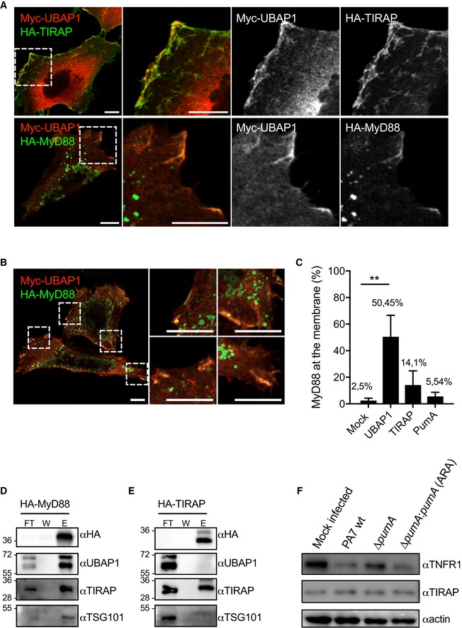
Figure 7. Analysis of the impact of UBAP1 on TIRAP and MyD88.

-
ARepresentative micrographs obtained by confocal microscopy of HeLa cells co‐expressing Myc‐UBAP1 (red) and adaptor proteins HA‐TIRAP (green, top panel) or HA‐MyD88 (green, bottom panel). Cells were fixed after 10 h of transfection. Scale bars correspond to 10 μm.
-
BDifferent zoomed images showing HA‐MyD88 (green) recruitment to the plasma membrane in the presence of Myc‐UBAP1 (red). Scale bars correspond to 10 μm.
-
CQuantification of plasma membrane localization of MyD88 in cells expressing MyD88 alone or with either UBAP1, TIRAP or PumA. At least 200 cells were enumerated in three independent experiments, and membrane localization was defined under the strict criteria of clear line at the plasma membrane. Cells with MyD88‐positive vesicles in close proximity to the plasma membrane were not counted as positive. Non‐parametric one‐way ANOVA Kruskal–Wallis test was performed, with Dunn's multiple comparisons test. **P < 0.01.
-
D, EEndogenous co‐IP from cells expressing (D) HA‐MyD88 and (E) HA‐TIRAP. The fractions bound to HA‐trapping beads were probed with anti‐HA, anti‐UBAP1, anti‐TIRAP and anti‐TSG101 antibodies. Non‐bound fraction (FT), last wash (W) and elution (E) are shown for each sample and the molecular weights indicated (kDa).
-
FWestern blot of TNFR1 in A549 cells infected for 1 h with either P. aeruginosa PA7 wt, ∆pumA or ∆pumA:pumA (Ara) induced with 1% arabinose. A mock‐infected sample was included as a negative control. The same blot was also probed for TIRAP and actin to control loading.
Source data are available online for this figure.
