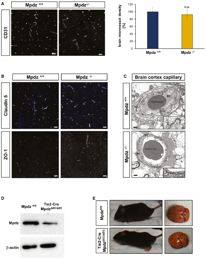
Figure EV1. Loss of Mpdz does not alter the brain microvasculature and endothelial‐restricted loss of Mpdz does not cause hydrocephalus.

- Brain sections from Mpdz −/− and littermate Mpdz +/+ mice were stained for the endothelial marker CD31. Scale bars, 50 μm. Graph showing relative changes in microvessel density. n = 3 animals per genotype; n.s., not significant (unpaired two‐sided Student's t‐test). Data are presented as mean ± SD.
- Brain section stained for the tight junction proteins claudin‐5 and ZO‐1. No differences were observed between Mpdz −/− and littermate Mpdz +/+ mice at P7. Scale bar, 50 μm.
- Transmission electron micrograph of cortical brain capillaries shows no difference in tight junction assembly between Mpdz −/− and Mpdz +/+ littermates. Boxes are highlighting endothelial cell junctions. One representative box shows the area as a zoom‐in. Scale bars, 1 μm and 100 nm. EC, endothelial cell; PC, pericyte.
- Western blot to determine Mpdz protein expression in isolated lung endothelial cells from adult Tie2‐Cre;Mpdz ΔEC/ΔEC mice.
- Endothelial cell‐specific Mpdz‐deficient mice (ΔEC/ΔEC) developed normally with no indication for hydrocephalus (n > 200 ΔEC/ΔEC observed for more than four months of age). H&E staining of brain sections from three‐month‐old mice.
Source data are available online for this figure.
