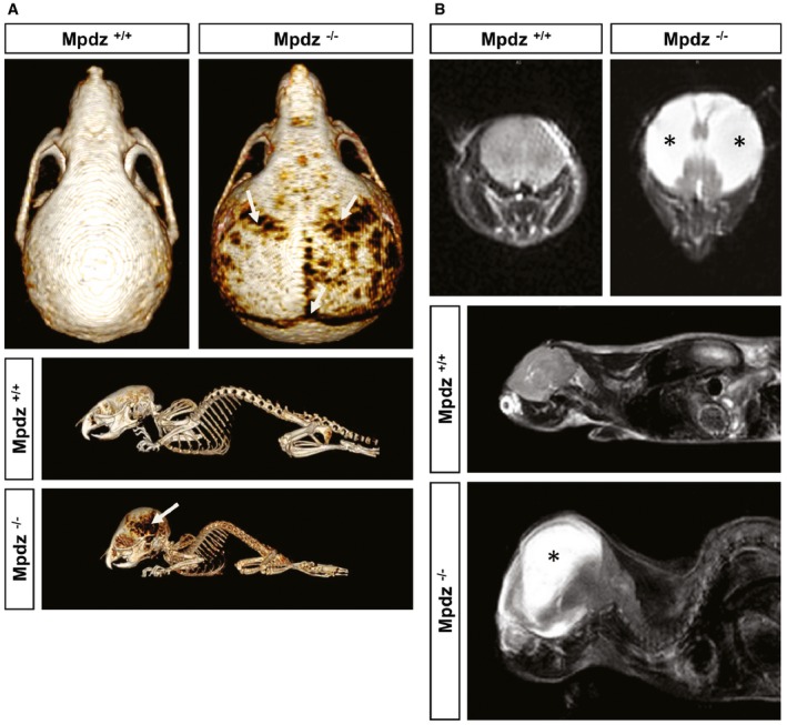
Figure 2. Mpdz‐deficient mice develop hydrocephalus.

- At postnatal day 27 (P27), Mpdz −/− and wild‐type littermates were subjected to a computed tomography. Three‐dimensional reconstruction shows macrocephaly and thinning of skull bones (arrows).
- T2‐weighted coronal and sagittal magnetic resonance images of the head of Mpdz −/− vs. Mpdz +/+ mice at P27. CSF in the enlarged lateral ventricles appears hyperintense (asterisks).
