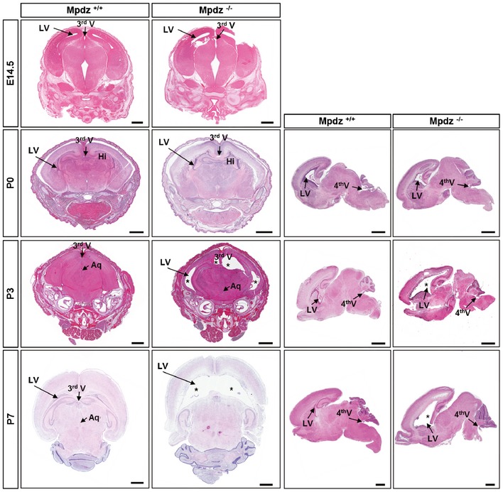
Figure 3. Mpdz −/− mice develop postnatal hydrocephalus.

H&E staining of brain sections from Mpdz −/− and littermate Mpdz +/+ mice at different developmental stages. At embryonic stage E14.5 and at birth (P0), coronal sections showed no overt alterations in Mpdz −/− mouse brains. At P3 and P7, enlarged lateral ventricles (*) were detected in Mpdz −/− mice. Horizontal sections demonstrate ventriculomegaly in Mpdz −/− mice at P3 and P7. Aq, cerebral aqueduct; Hi, hippocampus; LV, lateral ventricle; V, ventricle (3rd, 4th). Scale bars: E14.5, 500 μm; P0, P3, and P7, 1 mm.
