
Figure 4. Impaired flow of CSF through the cerebral aqueduct after postnatal day 3.
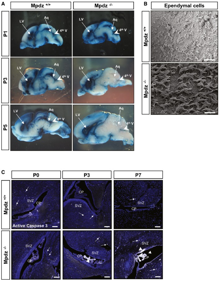
- Flow of Evans blue dye injected into a lateral ventricle trough the ventricular system of Mpdz −/− and Mpdz +/+ littermates was analyzed at postnatal day 1 (P1), P3, and P5. Sagittal sections show the lateral (LV) and fourth ventricles (4th V, arrowhead). In mice at P1, flow of Evans blue from one lateral ventricle through the third into the contralateral and the fourth ventricles was unremarkable in Mpdz −/− and Mpdz +/+ mice. At P3 and P5, no flow of Evans blue through the cerebral aqueduct (Aq) into the fourth ventricle could be detected in Mpdz −/− mice.
- Scanning electron micrograph of ependymal cells lining the roof of lateral ventricles at P7. No morphological alterations in motile cilia of Mpdz −/− compared to Mpdz +/+ mice. Scale bar, 20 μm.
- Antibody staining against cleaved caspase‐3 (white color, arrows and arrowheads) to detect apoptotic cells. CP, choroid plexus; SVZ, subventricular zone. Scale bar, 100 μm.