
Figure EV4. Human ES cell‐derived cardiomyocytes show circadian oscillation in Bmal1‐ and Per2‐dLuc.
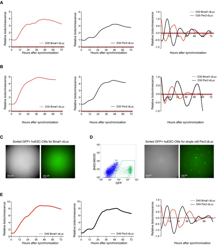
- Promoter‐based destabilized luciferase (dLuc) reporter assay of the Bmal1 (red) and Per2 (black) promoter in synchronized human ES cell‐derived cardiac cells at D30 (replicate for Fig 3D). Values are relative to T0. Right: Detrended Bmal1‐dLuc and Per2‐dLuc luciferase signal. Measurements were performed using a LumiCycle32.
- Similar analysis as in (A) on an additional independent replicate.
- Brightfield and fluorescent image of D30 GFP+ sorted human ES cell‐derived cardiomyocytes used for analysis in Fig 3F (left/red).
- Similar analysis as in (A) for D45 human ES cell‐derived cardiac cells.