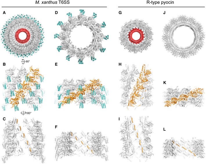
Figure 4. Comparing the models of Myxococcus xanthus T6SS sheath and R‐type pyocin in different conformations.

-
ATop view of the extended M. xanthus T6SS sheath model containing five layers of MxTssBC‐MxHcp hexamers. The sheath is colored gray, with the recycling domain colored cyan. The Hcp inner tube is colored red.
-
BSide view of the model in (A), with only the front half shown for clarity. One protofilament of the sheath is colored orange.
-
CBack view (from inside the tube) of model shown in (B). Here, only the attachment α‐helices in one protofilament of the sheath are colored orange.
-
D–FThe same representations as in (A–C) but for the contracted M. xanthus T6SS sheath.
-
G–LThe same representations as in (A–F) but for the R‐type pyocin.
