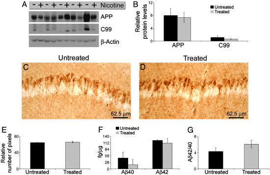
Fig. 3.
APP processing and Aβ deposition are not altered after chronic nicotine administration. (A) Immunoblot shows that APP and C99 levels are not significantly different between treated and untreated 3xTg-AD mice. (B) Quantitative analysis of blots in A after normalizing to β-actin shows that nicotine administration did not significantly alter the steady-state levels of APP or C99. (C and D) Immunohistochemical analysis by using an anti-Aβ42 specific antibody shows that Aβ deposition was not altered after chronic nicotine administration. (E) Densitometric analysis of C and D did not reveal any significant change in the Aβ load in the hippocampus of treated versus untreated 3xTg-AD mice. (F) Sandwich ELISA revealed that Aβ40 and Aβ42 steady-state levels were unaltered after chronic nicotine administration (n = 5 per group). Although there appears to be reduced Aβ40 levels in the treated mice, it did not achieve significance (P = 0.332 and 0.676 for Aβ40 and Aβ42, respectively). (G) The ratio of Aβ42/Aβ40 was also unchanged by the nicotine administration (P = 0.198).

