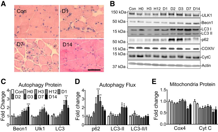
Fig. 3.
Autophagy-related protein content following hindlimb ischemia-reperfusion injury. A: representative histological images of regenerating plantaris muscle at 24 h and 7 and 14 days postinjury relative to uninjured control (Con): scale bar = 100 μm. B: representative immunoblot images of Ulk1, Beclin1, p62, LC3-I, LC3-II, Cyt C, COX4, and actin from the plantaris muscle at 0, 3, 12, 24, 48, and 72 h, and 7 and 14 days post-ischemia-reperfusion (I/R) injury and Con control. C: quantification of Beclin1, Ulk1, and total LC3, collectively an index of autophagy capacity, during muscle regeneration. D: quantification of p62, LC3-II collectively as an index of autophagy flux, during muscle regeneration. E: quantification of COX4 and Cyt C as a measure of mitochondrial content. Results are represented as means ± SE for n = 6 mice for each time point. *P < 0.05, **P < 0.01, ***P < 0.0001 significantly different from Con.

