Fig. 1.
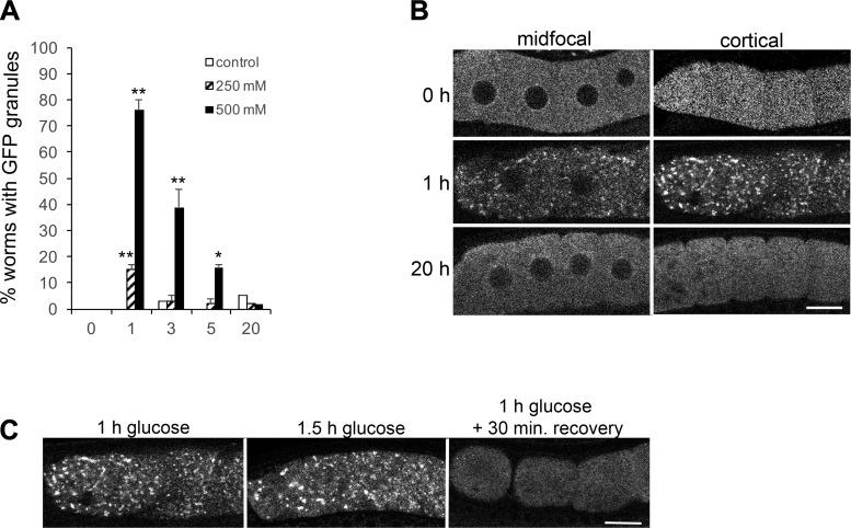
Short-term exposure to glucose induces the assembly of large GFP::MEX-3 granules in oocytes. A: 1-h exposure to 250 mM glucose induces the assembly of large GFP::MEX-3 granules in 15% of worms, but they are not maintained after 3, 5, or 20 h. Exposure to 500 mM glucose more robustly induces the assembly of large GFP::MEX-3 granules, but they are not maintained beyond 3 h of exposure. GFP granules were not detected in significant numbers of worms for any of the time-matched negative controls. Values are means ± SE (n = 89–101). **P < 0.001, compared with each time 0 control (for 250 or 500 mM glucose); *P < 0.01, compared with the time 0 control. B: live images of GFP::MEX-3 worms show the change in GFP distribution after 1 h of 500 mM glucose exposure, from being uniform throughout the cytoplasm at time 0, to becoming concentrated in discrete, cortical granules. The GFP granules are not maintained after 20 h of glucose exposure. Scale bar = 10 µm. C: GFP::MEX-3 granules dissociate within 30 min of recovery on nematode growth media plates. GFP::MEX-3 granules are still detected after 1.5 h of glucose exposure; thus the dissociation appears to be an active recovery process (n = 32–50). Scale bar = 10 µm.

