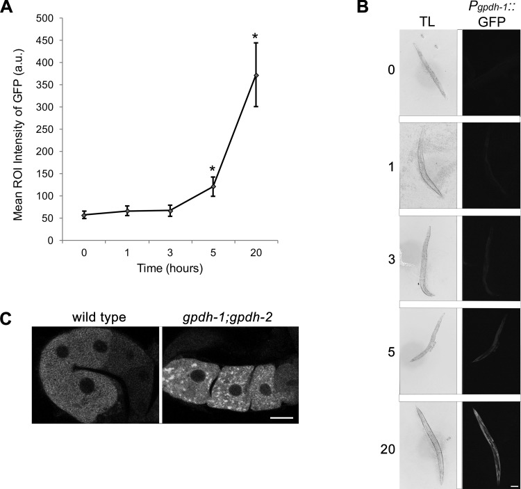
Fig. 4.
Inverse relationship between assembly of RNP granules and glycerol levels. A: levels of GFP in the Pgpdh-1::GFP strain increase significantly after 5 and 20 h of glucose exposure. Values are means ± SE (n = 27–49). *P < 0.001, compared with control. ROI, region of interest; a.u., arbitrary units. B: transmitted light (TL) and GFP fluorescence images of Pgpdh-1::GFP adults after 1, 3, 5, and 20 h of 500 mM glucose exposure. Scale bar = 100 µm. C: MEX-3 granules are detected in only 15% of wild-type worms after 5 h of glucose exposure. In contrast, MEX-3 granules are detected in 73% of gpdh-1;gpdh-2 worms after 5 h of glucose exposure (n = 17–28). Scale bar = 7 µm.

