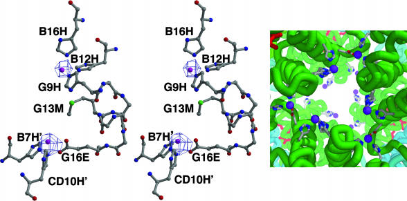
Fig. 2.
Zn2+-binding sites. (Left and Center) Stereoview of two Zn2+-binding sites with protein ligands along with a 2Fo – Fc map contoured at 6 σ. Each A2 subunit provides one complete Zn2+-binding site involving three His ligands and shares two others at interfaces with neighboring A2 subunits. The refined Zn2+ positions are shown as purple balls. (Right) The arrangement of Zn2+-binding sites near the molecular threefold axis of R. pachyptila C1 Hb. Zn2+ ions are purple, along with protein ligands and main-chain traces in the vicinity of the Zn2+ ions.

