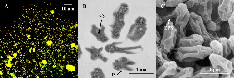
FIG 3.
Morphology of AM1. (A) Confocal laser scanning microscopy image of FISH-stained Nitrotoga cells (yellow) by Cy3-labeled NTG840, and other microorganisms (green) by fluorescein isothiocyanate (FITC)-labeled EUB338 mixture. (B) Overview of ultrathin sections of Nitrotoga-like bacteria observed by TEM. An extraordinarily wide periplasmic space (P) surrounded the cytoplasm (Cy). (C) SEM image of Nitrotoga-like bacterial cells loosely coupled by thin layers of extracellular polymeric substances (EPS).

