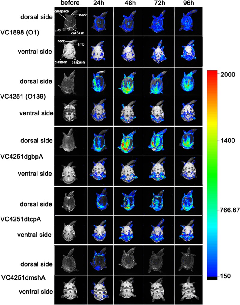
FIG 1.

Body surface bioluminescence imaging of soft-shelled turtles infected with V. cholerae strains. The soft-shelled turtles were separately immersed in bacterial suspensions of bioluminescence-labeled O1 strain VC1898, O139 strain VC4251, and three VC4251-derived gene mutant bioluminescent strains. All the dorsal and ventral body surfaces were scanned before infection (as negative controls) and at 24, 48, 72, and 96 h postinfection. The figure shows one representative turtle for each tested strain. The color bar on the right shows the intensity of bioluminescence, representing increasing densities of bacteria. The anatomical structures on the dorsal side are labeled in the upper left corner image.
