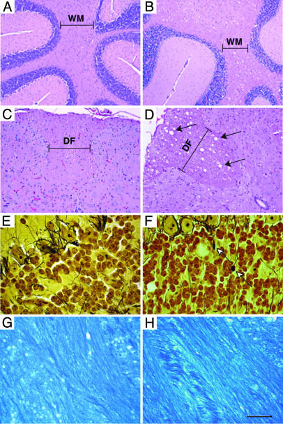
Fig. 3.
CNS pathology in Galgt1 Siat9 double-null mice. Tissue sections from WT (A, C, E, and G) and double-null (B, D, F, and H) mice are shown. (A and B) Hematoxylin/eosin-stained sections of the cerebellum from 2.5-month-old mice. Note the vacuoles in the white matter (WM) area of the double-null mice. (Bars: 100 μm.) (C and D) Hematoxylin/eosin-stained sections of the spinal cord from 5-month-old mice. Note the vacuoles (arrows) in the white-matter area (dorsal funiculus; DF) of the double-null mice. (Bars: 40 μm.) (E and F) Silver-stained sections of the cerebellum from 2.5-month-old mice. Note the axonal spheroids (arrows) in the double-null mice. (Bars: 20 μm.) (G and H) Luxol Fast Blue staining of the corpus callosum region shows that the degree of myelination is similar in WT and double-null mice. (Bars: 20 μm.)

