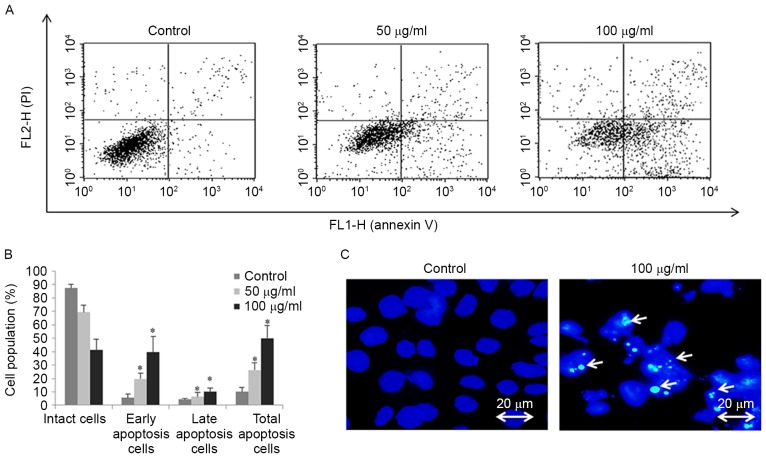
Figure 4.
FSB induces concentration-dependent apoptosis in AGS cells. AGS cells were treated with the indicated concentrations of FSB for 24 h. (A) Apoptosis was assessed using Annexin V-PI double staining and flow cytometry. (B) Histogram representation of apoptosis distribution of AGS cells treated with FSB. *P<0.05 vs. control. (C) AGS cells were stained with Hoechst 33,258 and analyzed using fluorescence microscopy (x 400). White arrows indicate bright blue regions which are fragmented or condensed nuclei. (FSB- flavonoid extract from Korean Scutellaria baicalensis Georgi; PI, propidium iodide).

