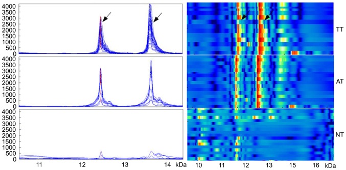
Figure 1.
The expression of two proteins in three different tissue types obtained from patients with Wilms tumor. Arrows denote the proteins at m/z12138 and m/z13462, identified as macrophage migration inhibitory factor and C-X-C motif ligand 7 chemokine, respectively. The left panel indicates simulated protein peaks, and the right panel of simulated electrophoresis reveals strong to weak expression (red to blue, respectively). TT, tumor tissue; AT, adjacent tissue; NT, normal tissue.

