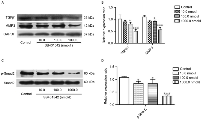
Figure 2.
RPMI 8226 cells were treated with SB431542 (10, 100 and 1,000 nmol/l) for 48 h. (A) Representative image and (B) quantification of expression of TGFβ1, MMP3 and GAPDH as determined by western blotting. (C) Representative image and (D) quantification of expression of Smad2 and p-Smad2 as determined by western blotting. Values are expressed as the mean ± standard deviation of 3 independent experiments. *P<0.05, ***P<0.001 vs. the control. GAPDH, glyceraldehyde-3-phosphate dehydrogenase; MMP3, matrix metallopeptidase 3; p, phosphorylated; Smad2, SMAD family member 2; TGFβ1, transforming growth factor β1.

