Figure 3.
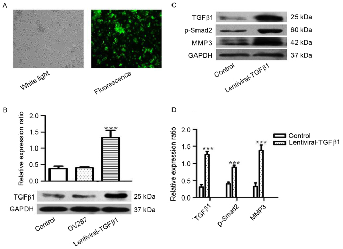
RPMI 8226 cells were transfected with lentiviral-TGFβ1 vectors and (A) the transfection efficiency was assessed by fluorescence microscopy. (B) The expression of TGFβ1 was assessed by western blotting in RPMI 8226 cells transfected with lentiviral-TGFβ1 vectors, GV287 and non-transfected cells (control). (C) The expression of TGFβ1, p-Smad2 and MMP3 was analyzed by western blotting in RPMI 8226 cells transfected with lentiviral-TGFβ1 vectors and in non-transfected cells (control). (D) The ratios of TGFβ1, p-Smad2 and MMP3 relative to GAPDH were calculated. Values are expressed as the mean ± standard deviation of 3 independent experiments. ***P<0.05 vs. the control. GAPDH, glyceraldehyde-3-phosphate dehydrogenase; MMP3, matrix metallopeptidase 3; p-Smad2, phosphorylated SMAD family member 2; TGFβ1, transforming growth factor β1.

