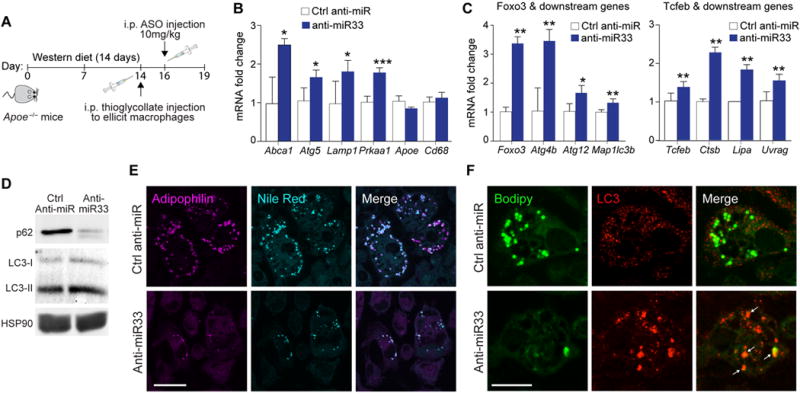
Figure 2. Inhibition of endogenous miR-33 promotes autophagy in macrophage foam cells in vivo.

A) Schematic diagram showing the experimental outline for antisense oligonucleotide treatment of male Apoe−/− mice on a Western diet. B-C) qPCR of (B) miR-33 target genes or non-miR-33 targets, as well as (C) Foxo3, Tcfeb and their downstream transcriptional targets in macrophage foam cells isolated hyperlipidemic Apoe−/− mice treated with anti-miR-33 or control anti-miR. D) Western blotting of lysates from peritoneal macrophages isolated from Apoe−/− mice treated with anti-miR-33 or control anti-miR to detect LC3-I (cytosolic), LC3-II (autophagosomal) or the autophagy-chaperone protein p62. HSP90 is shown as an internal control. E) Fluorescence microscopy imaging of Adipophilin and neutral lipid (Nile Red-positive) droplets in peritoneal macrophages isolated from hyperlipidemic Apoe−/− mice treated with anti-miR-33 or control anti-miR. Scale bar = 25μm. F) Fluorescence microscopy imaging of LC3 and neutral lipid (BODIPY-positive) droplets in peritoneal macrophages isolated from hyperlipidemic Apoe−/− mice treated with anti-miR-33 or control anti-miR. Scale bar = 25μm. Data are the mean ± s.e.m. of 3 mice. *P<0.05, **P<0.005.
