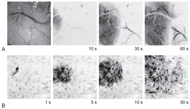
Fig. 7.1.
Cortical spreading depression (CSD) and astrocyte calcium waves. (A) Optical imaging of the cortical surface in a mouse. Far left panel shows image of mouse cortex visualized through the thinned skull of an anesthetized mouse (a recording electrode is seen in the upper right of the field; image scale = 1200 × 1200 μm). Subsequent images show change in reflectance of this area of cortex over time associated with a CSD wave. A change in optical signal of the parenchyma spreads slowly across the cortex. Dilation (darkening) of surface arteries propagates ahead of the CSD wavefront, followed by constriction of these vessels accompanying the CSD wave. (B) Astrocyte calcium wave. Images show fluorescence of the calcium indicator fluo-4 with an inverse gray scale (darker gray indicates higher intracellular calcium; image scale = 400 × 400 μm). Mechanical stimulation of a single cell with a micropipette evokes a wave of increased calcium concentration that spreads from cell to cell over as many as hundreds of cells. The temporal and spatial characteristics of this wave are remarkably similar to those of CSD.

