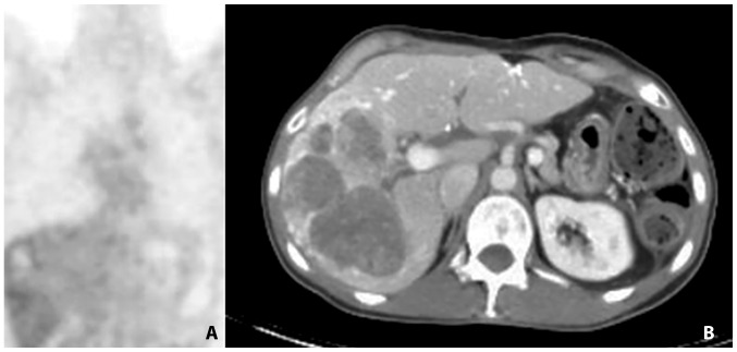
Figure 2.
18F-FDG PET-CT in March 2012. (A) Metabolic response (no 18FDG-uptake) on PET scan in 1 coronal plane image. (B) liver metastases on CT scan in 1 cross-sectional image. An increased volume of the liver metastases with minimal 18F-FDG uptake on PET suggests a metabolic response with necrosis. 18F-FDG, 18Fludeoxyglucose; PET-CT, positron emission tomography-computed tomography.

