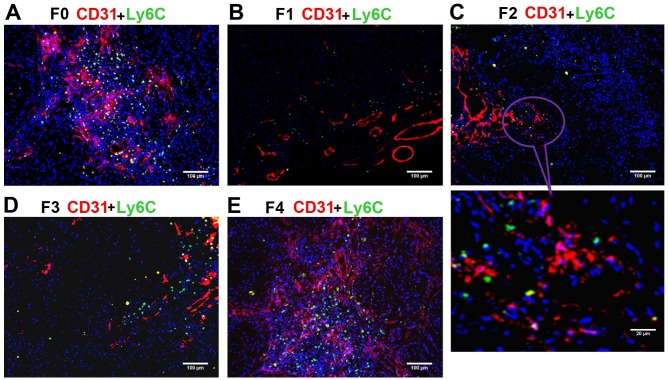
Figure 4.
Immunostaining of Ly6C+ cell subsets (green) and CD31 (red) in non-small cell lung cancer xenografts. Blue fluorescence indicates DAPI (nuclear) staining. (A) The F0 (control) group had a large percentage of Ly6C+ cells. (B) F1, (C) F2 and (D) F3 groups displayed a lower percentage of Ly6C+ compared with that in F0. (E) The percentage of Ly6C+ in the F4 group was similar to that in F0 and higher compared with that in F1, F2 and F3. Ly6C, lymphocyte antigen 6C; CD, cluster of differentiation; F0 mice, injected with A549 cells only; F1 mice, injected with A549 cells and treated with B20 twice weekly; F2 mice, transplanted with F1 tumor explant and treated with B20 twice weekly; F3 mice, transplanted with F2 tumor explant and treated with B20 twice weekly; F4 mice, transplanted with F3 tumor explant with no treatment.

