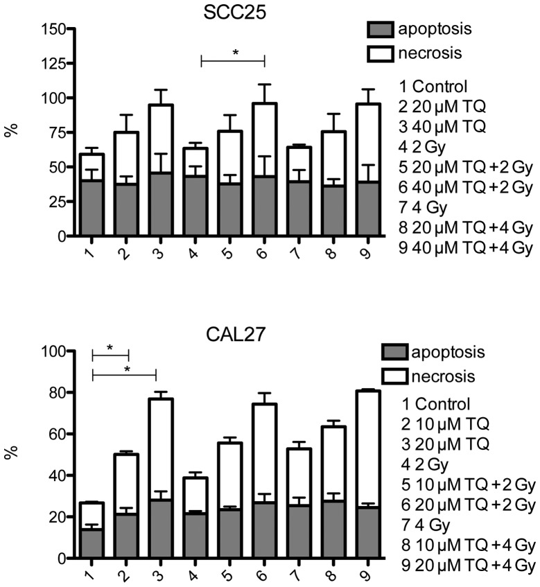
Figure 4.
Evaluation of apoptosis by flow cytometry. The cells were treated with thymoquinone (TQ) (SCC25 with 0, 40 and 60 µM; and CAL27 with 0, 20 and 40 µM). Apoptosis was measured after 48 h by flow cytometry using the Annexin-V Apoptosis Detection kit. *P=0.0021 for SCC25 cells treated with 40 µM TQ + 2 Gy vs. SCC25 cells treated with 2 Gy only, P=0.0640 for CAL27 cells treated with 10 µM TQ vs. control cells and P<0.0001 for CAL27 cells treated with 20 µM TQ vs. control cells.

