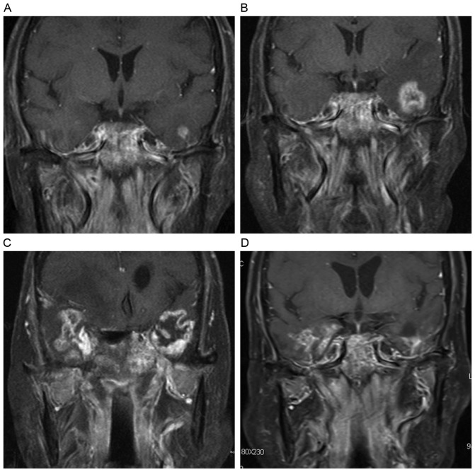
Figure 3.
Evolution of a contrast enhanced lesion. (A) A post-contrast coronal T1-weighted image reveals a solid enhanced nodular lesion with homogeneous hyperintensity in the left temporal lobe. (B) The lesion enlarged in size 4 months later, and presented as an enhanced nodular lesion with necrosis. (C) After 6 months, a finger-like enhanced lesion was observed. (D) A dotted and patchy enhanced lesion appeared 12 months later, along with temporal lobe atrophy.

