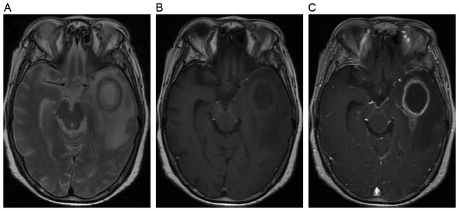
Figure 6.
Example of magnetic resonance images of a cyst lesion from a 55-year-old female patient. (A) Axial T2-weighted image shows an oval cyst with inner hyperintensity and outer annular hypointensity. (B) Axial T1-weighted image shows the oval cyst with inner hypointensity and outer annular hyperintensity. (C) Axial post-contrast T1-weighted image shows the cyst with annular enhancement.

