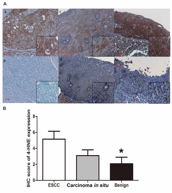
Figure 1.
Heterogeneous 4-HNE expression level in ESCC. (A) IHC analysis was performed on formalin-fixed paraffin-embedded tissues, and the expression levels of 4-HNE were investigated in the cytoplasm. Based on the staining, tumors in the cohort were categorized into (a) 4-HNE-positive or (b) 4-HNE-negative in ESCC tissues; the normal epithelium was categorized into (c) 4-HNE-positive or (d) 4-HNE-negative; carcinoma in situ was categorized into (e) 4-HNE-positive or (f) 4-HNE-negative. (B) IHC score of 4-HNE expression in ESCC tissues in situ and benign tissues. IHC staining scale bar, 50 µm. *P<0.05. 4-HNE, 4-hydroxynonenal; ESCC, esophageal squamous cell carcinoma; IHC, immunohistochemistry.

