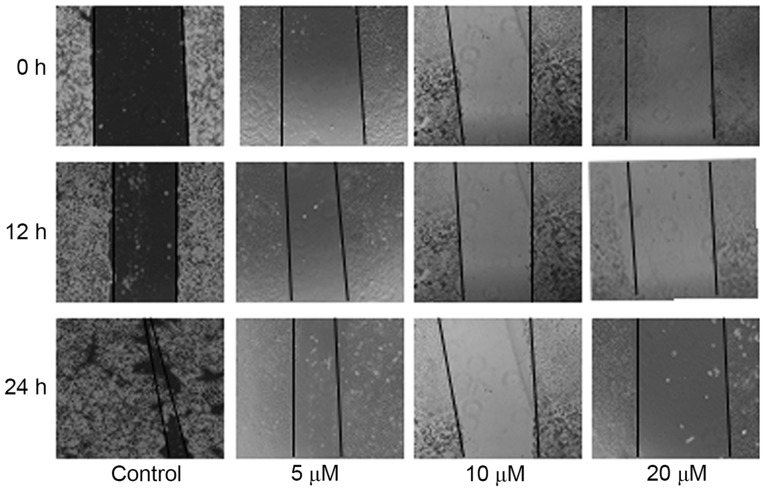
Figure 2.
Migration ability as evaluated by wound-healing assays. HepG2 cells were treated with 5, 10 or 20 µM SM, and the width of the wound exhibited a lower propensity for closure compared with the control group. The untreated HepG2 cells filled the majority of the wounded area after 24 h, whereas a distinct gap remained in the SM-treated groups. SM inhibited wound closure of the HepG2 cells in a dose-dependent manner. Magnification, ×40. SM, solamargine.

