
Figure 5.
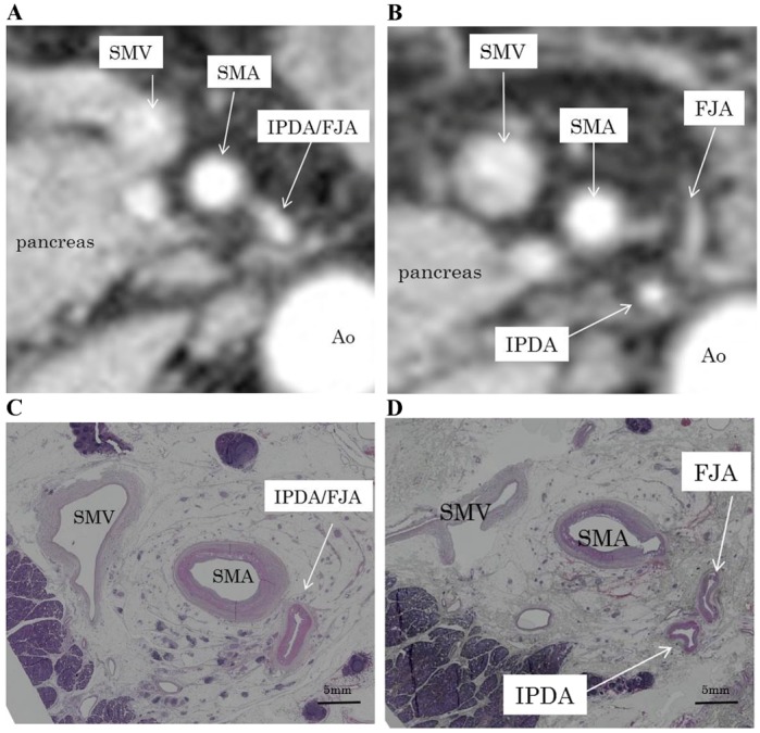
(A) CT scan showing the IPDA/FJA common vessel emerging between the left and dorsal sides of the SMA. (B) CT scan showing the IPDA branching from the FJA. (C) Surgical specimen showing the IPDA/FJA common vessel emerging between the left and dorsal sides of the SMA. (D) Surgical specimen showing the IPDA branching from the FJA. Magnification, ×1. CT, computed tomography; IPDA, inferior pancreaticoduodenal artery; FJA, first jejunal artery; SMA, superior mesenteric artery; SMV, superior mesenteric vein; Ao, aorta.