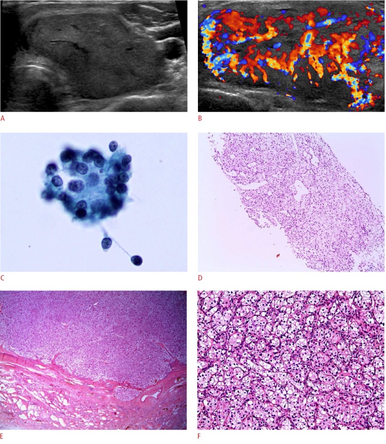
Figure 1. A 59-year-old woman with metastatic thyroid carcinoma (case 3).

A, B. The patient presented with a palpable neck mass that showed increased fluorodeoxyglucose uptake in a positron emission tomography scan performed 12 years after renal cell carcinoma was initially diagnosed. Ultrasonography shows (A, transverse; B, color Doppler) a 4.3×2.6×5.1-cm hypoechoic solid mass with a well-defined smooth margin and extensive vascularity. C. Fine-needle aspiration reveals a few round cell clusters showing atypia of undetermined significance/follicular lesion of undetermined significance (Papanicolaou, ×400). D. A core needle biopsy shows a solid trabecular tumor with clear cytoplasm (H&E, ×100). E. After a thyroidectomy, a well-demarcated expanding tumor with a fibrous capsule is shown in low magnification (H&E, ×12). F. In higher magnification, the tumor shows clear and eosinophilic cytoplasm (H&E, ×200).
