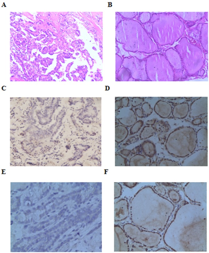
Figure 1.
Representative cell images. Hematoxylin and eosin staining for samples of (A) PTC and (B) normal thyroid tissues. Immunohistochemical photomicrographs of Beclin-1 and LC3-II in tissue samples from PTC. Negative (C) Beclin-1 and (E) LC3-II immunostaining in PTC tissues. Positive (D) Beclin-1 and (F) LC3-II immunoreactivity in normal cells. All images show magnification, ×200. PTC, papillary thyroid carcinoma; LC3-11, microtubule-associated protein 1 light chain 3.

