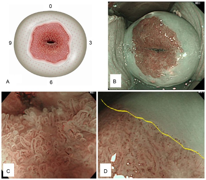
Figure 1.
Normal uterine cervix. (A) Schema of a normal uterine cervix; internal ostium of the uterus; columnar, transitional and squamous epithelium (in order from the inside). (B) Distant view of cervix using NBI mode. (C) NBI-ME image of normal columnar epithelium, with villous-like structures. (D) NBI-ME image of normal transitional epithelium (inside the yellow line), with a low height structure and NBI-ME image of normal squamous epithelium (outside the yellow line) with absence of structure. NBI-ME, narrow band imaging with magnification endoscopy.

