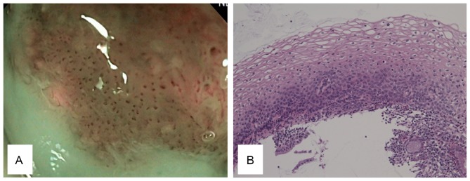
Figure 3.
CIN2 morphology. (A) CIN2 tumors present with a mild increase in the number of dots compared with the normal site, with no irregular arrangements. (B) The histological findings of CIN2 tumors include the presence of dysplastic cells in the basal two-thirds of the epithelium, and differentiation and maturation in the upper half of the epithelium (magnification, ×20). CIN, cervical intraepithelial neoplasia.

