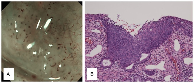
Figure 5.
CIS morphology. (A) CIS presents with elongated dots with severely irregular arrangements and a large increase in the number of dots compared with the normal site. (B) The histological findings of CIS include neoplastic cells from the base to the surface with cytological atypia, bristle mitoses and vascular hyperplasia (magnification, ×20). CIS, carcinoma in situ.

