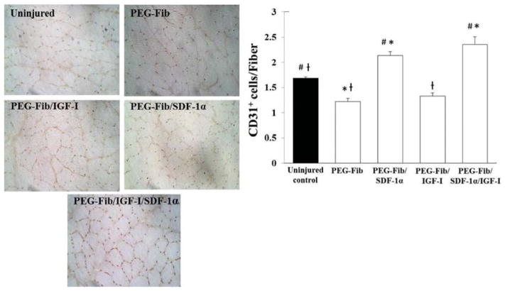
Fig. 3.
Identification and quantification of CD31+ cells within I/R injured skeletal muscle 14 days post-reperfusion. Animals were treated with PEGylated fibrin (PEG-Fib), PEGylated fibrin conjugated to SDF-1α (PEG-Fib/SDF-1α), PEGylated fibrin conjugated to IGF-I (PEG-Fib/IGF-1), and PEGylated fibrin conjugated to SDF-1α and IGF-I (PEG-Fib/SDF-1α/IGF-I) 24 h after TK-I/R injury and analyzed 14 days post-reperfusion. Representative images of CD31+ staining (200×) and quantification of CD31+ cells/muscle fiber (n = 3, 3 fields of view per animal). Values expressed as mean ± SEM, oneway ANOVA, Tukey post-hoc: *p < 0.05 versus uninjured control, #p < 0.05 versus PEG-Fib group, †p < 0.05 versus PEG-Fib/SDF-1α group.

