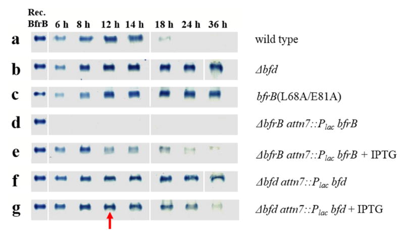
Fig. 4.

Monitoring the deposition and subsequent mobilization of iron in BfrB using native PAGE and Ferene-S staining. Wild type and mutant cells were cultured in PI media supplemented with 10 μM Fe. (a) Wild type cells deposit iron in BfrB during late exponential and early stationary growth phases, which are subsequently mobilized in late stationary phase. In contrast, the deposition of iron in BfrB in cultures of (b) Δbfd and (c) bfrB(L68A/E81A) cells is irreversible. (d) ΔbfrB cells do not accumulate iron, but (e) complementing ΔbfrB cells by expressing BfrB from an IPTG-inducible bfrB gene inserted into neutral site in the genome restores iron accumulation in BfrB followed by its mobilization in late stationary phase. (f) Δbfd cells accumulate iron irreversibly in BfrB, but (g) addition of IPTG at 12 h post inoculation (red arrow) induces the expression of Bfd from an IPTG-inducible bfd gene inserted into a neutral site of the genome and promotes iron mobilization from BfrB).
