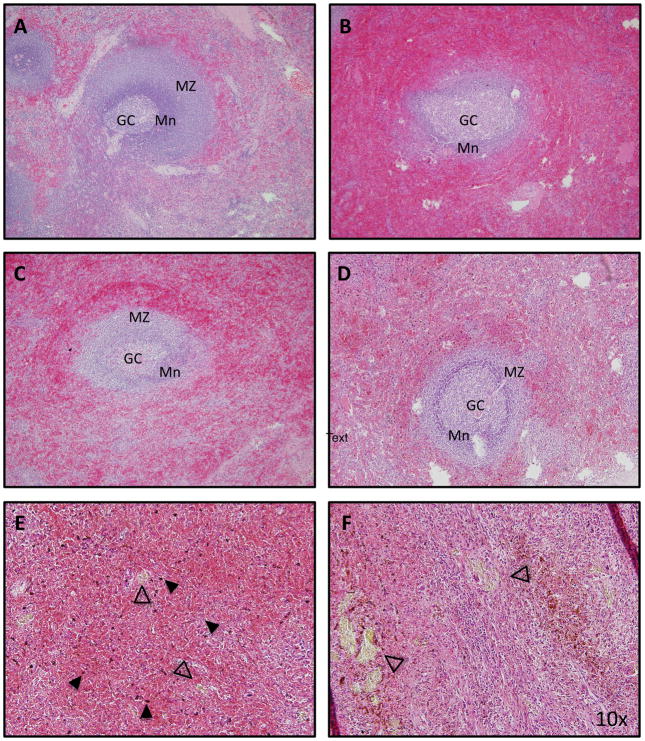
Figure 2. Histopathology of the spleen from patients with sickle cell disease.
(A) In normal spleen tissue, the follicle is organized into germinal centre (GC), mantle zone (Mn) and marginal zone (MZ).Senescent red cells are trapped and destroyed in the surrounding red pulp. (B) In a US patient with SCD who was splenectomised following recovery from splenic sequestration, the red pulp is congested with sickled cells. The structure of the follicle is less well defined. (C) An African patient with SCD who was splenectomised for splenic sequestration demonstrates congested of red pulp and preserved follicular architecture. (D) An African SCD patient who was splenectomised for hypersplenism similarly demonstrates engulfment of red pulp and preserved follicular architecture. In this patient, haemozoin deposits (E, solid arrowhead) and gamna gandy bodies are prominent (E and F, open arrowhead). All images are stained with haematoxylin and eosin stain. Magnification: 10x.

