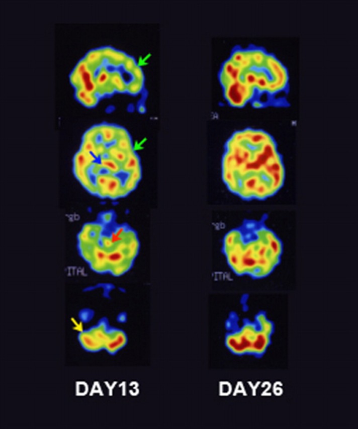
Figure 1.

123I-iodoamphetamine single photon emission CT (123I-IMP SPECT) imaging of the patient. On day 13, 123I-IMP SPECT reveals decreased rCBF in the frontotemporal cortex (green arrows), thalamus (blue arrow), brainstem (red arrow) and cerebellum (yellow arrow) (A). Follow-up 123I-IMP SPECT on day 26 shows improvement in the damaged lesion (B).
