Fig. 2.

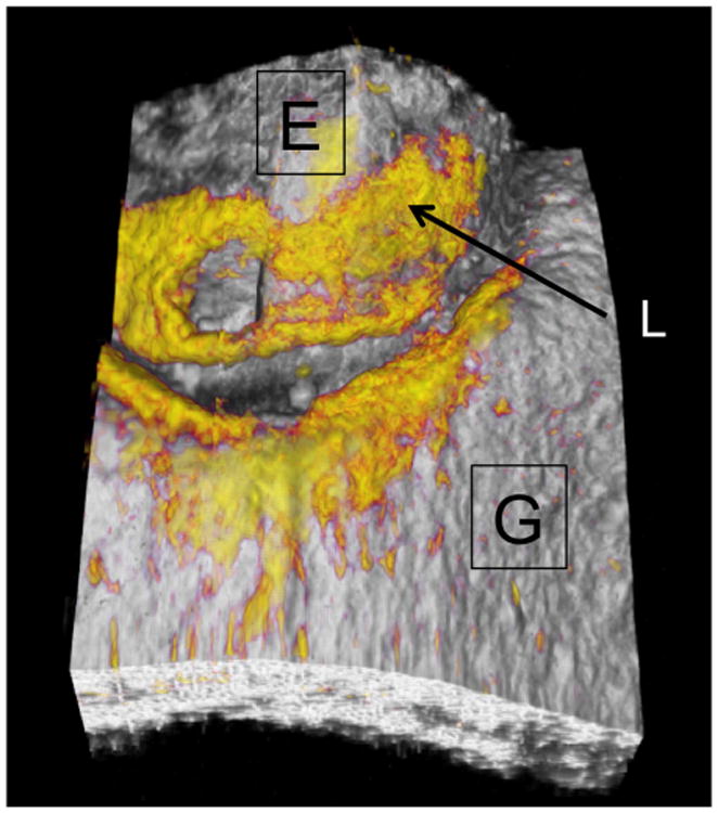
A surface rendering of a typical 3D image acquired showing the position of the enamel (E) and the gingiva (G) with the areas of high reflectivity shown in yellow. High reflectivity areas on the enamel indicate the position of the lesion (L).

A surface rendering of a typical 3D image acquired showing the position of the enamel (E) and the gingiva (G) with the areas of high reflectivity shown in yellow. High reflectivity areas on the enamel indicate the position of the lesion (L).