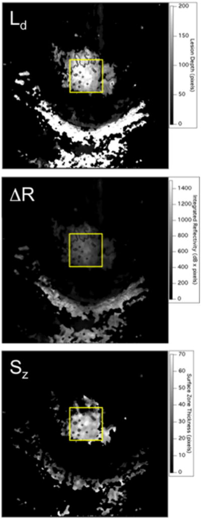
Fig. 3.

Two dimensional projection images of ΔR, Ld, and Sz, calculated for one of the lesions at a single time point. The lesion is located in the area of the yellow rectangular box. The half moon shaped region of high reflectivity below the yellow box is the gingiva.
