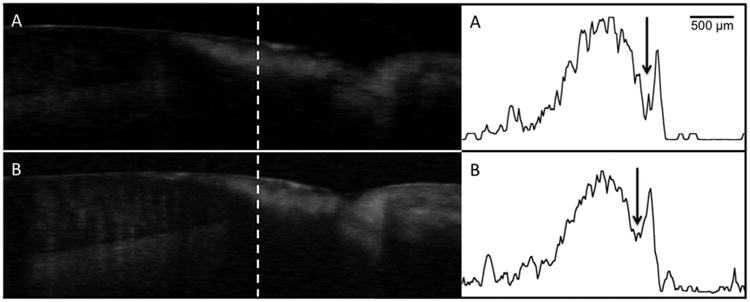
Fig. 4.

Processed CP-OCT b-scans of a cervical enamel lesion taken at week 0 (A) and again at week 30 (B). The lesion is clearly visible and it has a well-defined surface zone (Sz) that is visible. The dentinal enamel junction (DEJ) and the gingival (G) are visible in the image and the position of the scans are indicated on the photograph of the tooth. A-scans extracted at the position of the dashed line from each image are shown on the right with the tooth surface oriented on the right. The weakly scattering surface zone is located at the position of the arrow. The sharp spike to the right of the arrow is reflection from the tooth surface and the lesion body is the large broad peak to the left of the arrow. There was little change in the lesion structure after 30-weeks.
