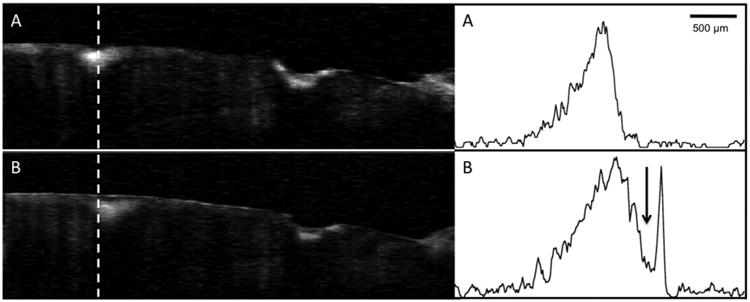
Fig. 6.

Processed CP-OCT b-scans of a tooth with two lesions acquired at week 0 and week 30. A-scans extracted at the position of the dashed line from each image are also shown. The surface zone thickness increased by 30% after 30-weeks. Note a well defined surface zone is also present on the cavitated lesion on the right as well as after 30-weeks.
